Primatologist

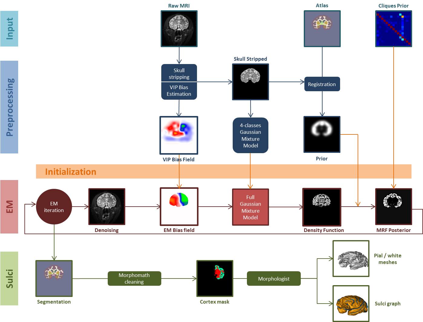
Primatologist is the main entry point of the Primatologist toolbox. It is a pipeline, i.e. a sequence of basic processes. Primatologist performs atlas registration, tissue segmentation and surfaces and sulci extraction. Its building blocks (the individual processing steps) are stored in blocks.

To this day, only the segmentation of anatomical regions has been thorouglly validated. Structures volume can be computed from a posteriori probability maps generated by the Primatologist pipeline with the hierarchical analysis process.

To process data, it is advised to use BrainVISA's databasing system. The usual practice is to create one database per study. Raw MR images should be imported with the dedicated importation process.

Description

Pipeline steps

Figure 1. The Primatologist pipeline.

Preprocessing and Atlas Registration

  1. Prepare MRI: select anatomical landmarks to orient correctly the input volume.
  2. (Align & Resample): use landmarks to align the brain and resample it at a fixed resolution.
  3. VIP Bias Correction: non-statistical bias field estimation.
  4. Skull Stripping: compute a mask of the intracranial volume.
  5. Select Evaluation Points: select randomly a subset of points for the affine transform estimation.
  6. Atlas to MRI: Affine Transform Estimation
  7. Atlas to MRI: Non-Linear Transform Estimation
  8. Atlas to MRI: Apply Transforms

Bayesian Segmentation

  1. Mixture Model Initialization: probabilizes the atlas if needed and initializes the GMM parameters.
  2. EM Segmentation: fits a statistical model to the MRI by expectation-maximization. Intensities are assumed to originate from a gaussian mixture model on a MRF lattice with non-stationary priors. This step also includes densoising and bias field estimation .

Surface Extraction and Sulci Segmentation

Convert Segmentation

  1. Compute Brain and Hemispheres Mask: includes some morphomathematical processing.
  2. Compute Cortex Mask: includes some morphomathematical processing.
  3. Histogram Analysis: performs a morphologist-compliant histogram analysis.
  4. (Invert T2 MRI): if the input MRI is T2-weighted, inverts it to make it T1-like (Morphologist only works with T1-weighted images).
  5. Talairach Transform Estimation: computes a transformation to Talairach space (mandatory for Morphologist processing).
Surfaces and Sulci Segmentation
  1. Topological Transform of the Cortex: fills the cortex mask towards the intracranial space boundaries.
  2. White Matter Surface Extraction.
  3. Skeletonization: skeletonizes the topologically transformed cortex to segment the sulci.
  4. Pial Surface Extraction.
  5. Cortical Folds Graph: extracts the sulci graph.

Provided Data

Macaque

CIVM Simplified

The Center for In Vivo Microscopy, in Duke University, has published a high resolution, post mortem atlas of the Rhesus Macaque (M. mulatta) brain. It contains anatomical (T2-weighted) and diffusion templates as well as a parcellation of the brain into 241 anatomic and functional structures.

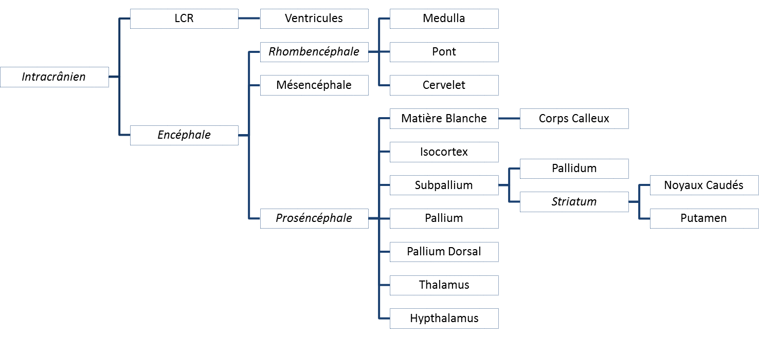
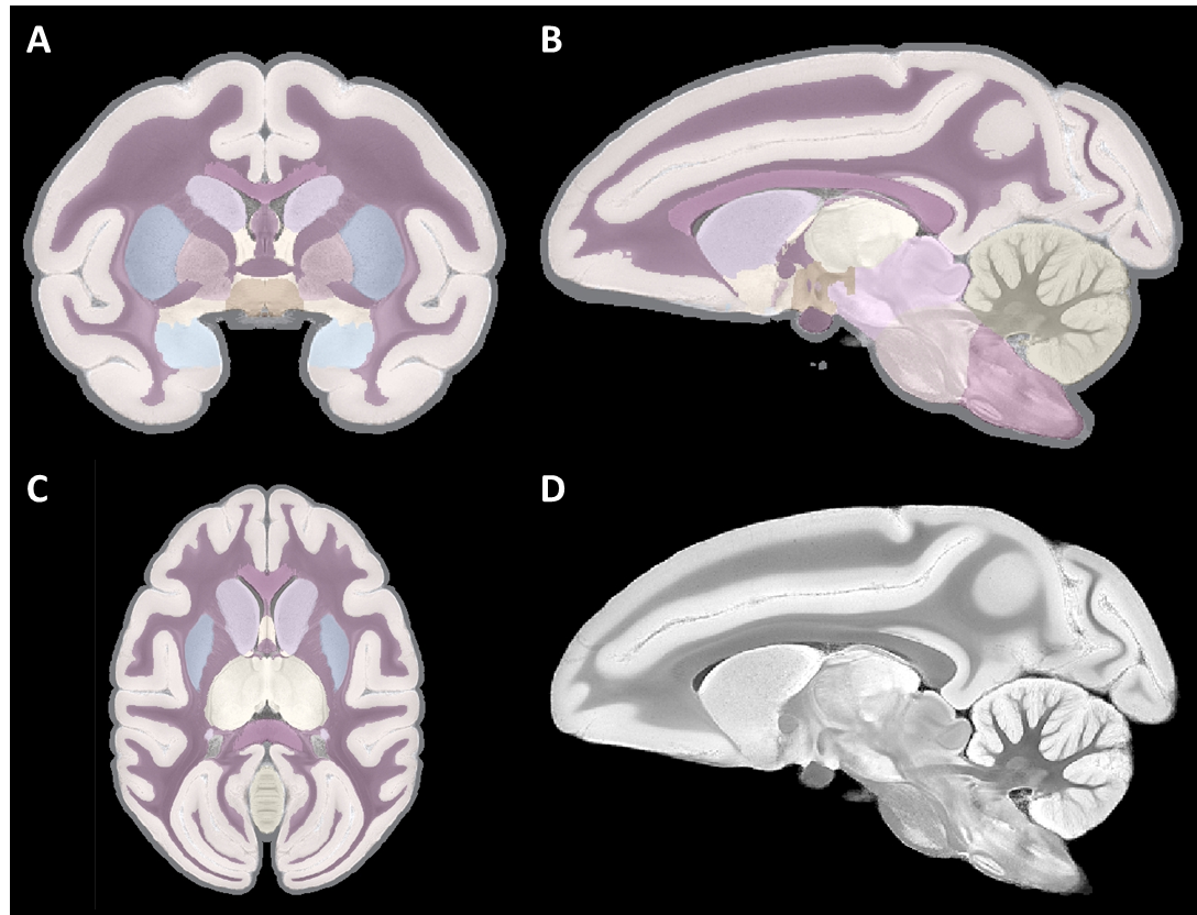
We have added to this parcellation two missing regions : a pseudo-CSF region was obtained by a 1mm dilation of the brain, and corpus callosum was manually segmented in sagittal incidence by a single operator. We then used our Atlas Hierarchy Tools to extract a subset of nodes (Figure 2), corresponding in the end to only 17 labels (Figure 3).

Figure 2. Selected nodes from the complete hierarchy. Pure virtual classes (i.e. without a label) are written in italic.
Figure 3. CIVM template and 17 selected labels.

Pseudo prior probabilities were obtained by extracting each label mask and smoothing it with a Gaussian kernel. Several of such probabilistic atlases are provided, with different smoothing radius. This task can also be performed during the segmentation process (see Basic Mixture).

This atlas has a very fine resolution (0.150 mm). We have thus also generated templates, labels and priors at coarser scales (0.3 and 0.6 mm). When using the optional resampling step (Align and Resmple), the MRI resampling and atlas resolutions should be the same.

To be usable with Primatologist, an atlas should also contain a hierarchy that associates labels with normalized names, another hierarchy that classifies structures between grey and white matters and CSF, and a Clique matrix that contains conditional neighbouring probabilities and which can be obtained from a volume of labels (see Compute Cliques from Labels).

Note:

For now, the naming norm is not described anywhere. To create your own Primatologist-compatible atlas, you should copy what was done for the CIVM atlas.

Parameters

input_mri: P:MRI Raw ( input )
specie: OpenChoice ( input )
contrast: OpenChoice ( input )
is_mapping: Boolean ( input )
point_anterior_commissure: Point3D ( input )
Anterior commissure point. This should be AC's most posterior points that intersects the interhemispheric plane.
point_posterior_commissure: Point3D ( input )
Posterior commissure point. This should be PC's most anterior points that intersects the interhemispheric plane.
point_interhemispheric: Point3D ( input )
Any point from the interhemispheric plane located above (i.e. dorsal to) the AC-PC line.
point_left_hemisphere: Point3D ( optional, input )
Any point located in the left hemisphere. If the MRI is in radiological orientation, this point should be located, on the screen, at the right of the interhemispheric plane.
template: P:Atlas Raw Template ( input )
labels: P:Atlas Raw Labels ( optional, input )
prior: P:Atlas Raw Prior ( optional, input )
lateralization: P:Atlas Raw Lateralization ( input )
clique_matrix: P:Atlas Clique Matrix ( input )
hierarchy: P:Atlas Labels Hierarchy ( input )
graywhite_hierarchy: P:Atlas Grey/White Hierarchy ( input )
output_atlas_labels: P:Atlas Registered Labels ( output )
output_em_posterior: P:EM Parcellation Proba ( output )
output_em_labels: P:EM Parcellation ( output )
output_greywhite_mesh_left: Hemisphere White Mesh ( output )
output_pial_mesh_left: Hemisphere Mesh ( output )
output_sulci_left: Cortical folds graph ( output )
output_greywhite_mesh_right: Hemisphere White Mesh ( output )
output_pial_mesh_right: Hemisphere Mesh ( output )
output_sulci_right: Cortical folds graph ( output )

Technical information

Toolbox : Primatologist

User level : 0

Identifier : Primatologist

File name : brainvisa/toolboxes/primatologist/processes/pipelines/Primatologist.py

Supported file formats :

input_mri :
gz compressed NIFTI-1 image, Aperio svs, BMP image, DICOM image, Directory, ECAT i image, ECAT v image, FDF image, FreesurferMGH, FreesurferMGZ, GIF image, GIS image, Hamamatsu ndpi, Hamamatsu vms, Hamamatsu vmu, JPEG image, Leica scn, MINC image, NIFTI-1 image, PBM image, PGM image, PNG image, PPM image, SPM image, Sakura svslide, TIFF image, TIFF image, TIFF(.tif) image, TIFF(.tif) image, VIDA image, Ventana bif, XBM image, XPM image, Zeiss czi, gz compressed MINC image, gz compressed NIFTI-1 image
template :
gz compressed NIFTI-1 image, Aperio svs, BMP image, DICOM image, Directory, ECAT i image, ECAT v image, FDF image, FreesurferMGH, FreesurferMGZ, GIF image, GIS image, Hamamatsu ndpi, Hamamatsu vms, Hamamatsu vmu, JPEG image, Leica scn, MINC image, NIFTI-1 image, PBM image, PGM image, PNG image, PPM image, SPM image, Sakura svslide, TIFF image, TIFF image, TIFF(.tif) image, TIFF(.tif) image, VIDA image, Ventana bif, XBM image, XPM image, Zeiss czi, gz compressed MINC image, gz compressed NIFTI-1 image
labels :
gz compressed NIFTI-1 image, Aperio svs, BMP image, DICOM image, Directory, ECAT i image, ECAT v image, FDF image, FreesurferMGH, FreesurferMGZ, GIF image, GIS image, Hamamatsu ndpi, Hamamatsu vms, Hamamatsu vmu, JPEG image, Leica scn, MINC image, NIFTI-1 image, PBM image, PGM image, PNG image, PPM image, SPM image, Sakura svslide, TIFF image, TIFF image, TIFF(.tif) image, TIFF(.tif) image, VIDA image, Ventana bif, XBM image, XPM image, Zeiss czi, gz compressed MINC image, gz compressed NIFTI-1 image
prior :
gz compressed NIFTI-1 image, Aperio svs, BMP image, DICOM image, Directory, ECAT i image, ECAT v image, FDF image, FreesurferMGH, FreesurferMGZ, GIF image, GIS image, Hamamatsu ndpi, Hamamatsu vms, Hamamatsu vmu, JPEG image, Leica scn, MINC image, NIFTI-1 image, PBM image, PGM image, PNG image, PPM image, SPM image, Sakura svslide, TIFF image, TIFF image, TIFF(.tif) image, TIFF(.tif) image, VIDA image, Ventana bif, XBM image, XPM image, Zeiss czi, gz compressed MINC image, gz compressed NIFTI-1 image
lateralization :
gz compressed NIFTI-1 image, Aperio svs, BMP image, DICOM image, Directory, ECAT i image, ECAT v image, FDF image, FreesurferMGH, FreesurferMGZ, GIF image, GIS image, Hamamatsu ndpi, Hamamatsu vms, Hamamatsu vmu, JPEG image, Leica scn, MINC image, NIFTI-1 image, PBM image, PGM image, PNG image, PPM image, SPM image, Sakura svslide, TIFF image, TIFF image, TIFF(.tif) image, TIFF(.tif) image, VIDA image, Ventana bif, XBM image, XPM image, Zeiss czi, gz compressed MINC image, gz compressed NIFTI-1 image
clique_matrix :
gz compressed NIFTI-1 image, Aperio svs, BMP image, DICOM image, Directory, ECAT i image, ECAT v image, FDF image, FreesurferMGH, FreesurferMGZ, GIF image, GIS image, Hamamatsu ndpi, Hamamatsu vms, Hamamatsu vmu, JPEG image, Leica scn, MINC image, NIFTI-1 image, PBM image, PGM image, PNG image, PPM image, SPM image, Sakura svslide, TIFF image, TIFF image, TIFF(.tif) image, TIFF(.tif) image, VIDA image, Ventana bif, XBM image, XPM image, Zeiss czi, gz compressed MINC image, gz compressed NIFTI-1 image
hierarchy :
Hierarchy, Hierarchy
graywhite_hierarchy :
Hierarchy, Hierarchy
output_atlas_labels :
gz compressed NIFTI-1 image, BMP image, DICOM image, Directory, ECAT i image, ECAT v image, FDF image, GIF image, GIS image, JPEG image, MINC image, NIFTI-1 image, PBM image, PGM image, PNG image, PPM image, SPM image, TIFF image, TIFF(.tif) image, VIDA image, XBM image, XPM image, gz compressed MINC image, gz compressed NIFTI-1 image
output_em_posterior :
gz compressed NIFTI-1 image, BMP image, DICOM image, Directory, ECAT i image, ECAT v image, FDF image, GIF image, GIS image, JPEG image, MINC image, NIFTI-1 image, PBM image, PGM image, PNG image, PPM image, SPM image, TIFF image, TIFF(.tif) image, VIDA image, XBM image, XPM image, gz compressed MINC image, gz compressed NIFTI-1 image
output_em_labels :
gz compressed NIFTI-1 image, BMP image, DICOM image, Directory, ECAT i image, ECAT v image, FDF image, GIF image, GIS image, JPEG image, MINC image, NIFTI-1 image, PBM image, PGM image, PNG image, PPM image, SPM image, TIFF image, TIFF(.tif) image, VIDA image, XBM image, XPM image, gz compressed MINC image, gz compressed NIFTI-1 image
output_greywhite_mesh_left :
GIFTI file, GIFTI file, MESH mesh, MNI OBJ mesh, PLY mesh, TRI mesh
output_pial_mesh_left :
GIFTI file, GIFTI file, MESH mesh, MNI OBJ mesh, PLY mesh, TRI mesh
output_sulci_left :
Graph and data, Graph and data
output_greywhite_mesh_right :
GIFTI file, GIFTI file, MESH mesh, MNI OBJ mesh, PLY mesh, TRI mesh
output_pial_mesh_right :
GIFTI file, GIFTI file, MESH mesh, MNI OBJ mesh, PLY mesh, TRI mesh
output_sulci_right :
Graph and data, Graph and data