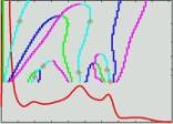
histo_analysis: Analyse d'histogramme ( entrée )
use_hfiltered: Booléen ( input )
hfiltered: T1 MRI Filtered For Histo ( optional, entrée )
use_wridges: Booléen ( input )
white_ridges: T1 MRI White Matter Ridges ( optional, entrée )
Toolbox : Morphologist
Niveau d'utilisateur : 0
Identifiant :
BrainvisaShowHistoAnalysisNom de fichier :
brainvisa/toolboxes/morphologist/processes/viewers/Segmentation/BrainvisaShowHistoAnalysis.pySupported file formats :
histo_analysis :Analyse d'histogramme, Analyse d'histogrammehfiltered :gz compressed NIFTI-1 image, Aperio svs, DICOM image, Répertoire, ECAT i image, ECAT v image, FDF image, FreesurferMGH, FreesurferMGZ, GIS image, Hamamatsu ndpi, Hamamatsu vms, Hamamatsu vmu, JPEG image, Leica scn, MINC image, NIFTI-1 image, SPM image, Sakura svslide, TIFF image, TIFF image, TIFF(.tif) image, TIFF(.tif) image, Ventana bif, Zeiss czi, gz compressed MINC image, gz compressed NIFTI-1 imagewhite_ridges :gz compressed NIFTI-1 image, Aperio svs, DICOM image, Répertoire, ECAT i image, ECAT v image, FDF image, FreesurferMGH, FreesurferMGZ, GIS image, Hamamatsu ndpi, Hamamatsu vms, Hamamatsu vmu, JPEG image, Leica scn, MINC image, NIFTI-1 image, SPM image, Sakura svslide, TIFF image, TIFF image, TIFF(.tif) image, TIFF(.tif) image, Ventana bif, Zeiss czi, gz compressed MINC image, gz compressed NIFTI-1 image