Select landmark points to accurately orient the MR volume.
Landmarks are :
the anterior commissure (AC)
the posterior commissure (PC)
a third point located in the interhemispheric plane (IHP), above the AC-PC line
a point located at the anatomical left of the brain
Scanners usually output volumes in the anatomical orientation, i.e. as if we were facing the subject. In this case, the anatomical left is located at the right of the screen.
Description
How to
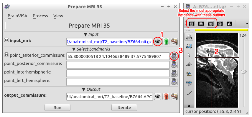
Open the input_mri in Anatomist by clicking the corresponding eye button ;
Locate the landmark (let's say the anterior commissure) in the image and click on it ;
Send the selected point coordinates to the landmark field (point_anterior_commissure) by clicking on the corresponding anatomist button ;
Repeat the above steps for the other landmarks.
Figure 1. Landmark selection steps.
Landmark position
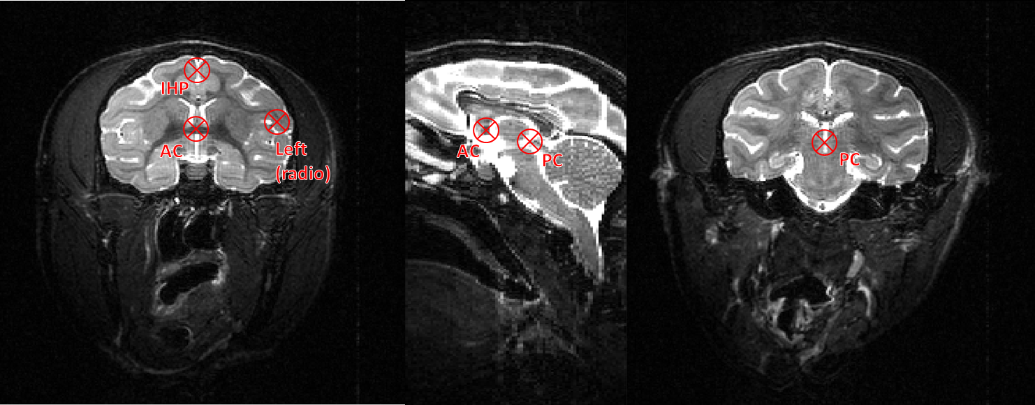
The position of points AC and PC is depicted Figure 2. The easiest way to find them is from the sagittal view: the user should find the intersection between each of these points and the interhemispheric plane. In the example image, because of the use of a stereotaxic frame, the interhemispheric plane is already aligned and both AC and PC can be selectedc from the same sagittal section. This might not always be the case though.
The interhemispheric point should be selected above the AC-PC line in order to correctly orient the volume.
If the scanner outputs image in radiological orientation, the left hemisphere point will be located, as in Figure 2, in the right side of the image. If no left hemisphere point is provided, the orientation will be kept as is. Else, the process will output the image in radiological orientation.
Figure 2. Location of AC and PC in the Macaque brain. The IHP should be selected above the AC-PC line.
Any point located in the left hemisphere. If the MRI is in radiological orientation, this point should be located, on the screen, at the right of the interhemispheric plane.
;
;