Morphologist
Brain segmentation and sulcal analysis
Fischer, G. Operto, S. Laguitton, M. Perrot, I. Denghien, D. Rivière, and J.-F. Mangin: Morphologist 2012: the new morphological pipeline of BrainVISA, In Proc. HBM, 2012. link bibtex-entry
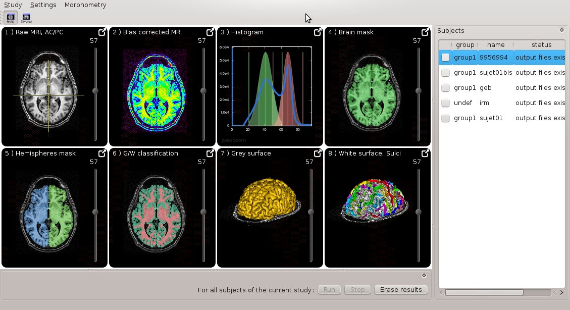
Morphologist UI
Morphologist now has a new graphical interface, featuring simplified user experience and quick interactive visualization:
Docmentation can be found here
It can be run either as a standalone program (morphologist), or through BrainVISA.
Morphologist toolbox in Brainvisa
Morpho-DeepSulci
SulPatEd
Sulpated, “Sulcal Patterns Editor” is a graphical interface to manually label sulci and sulcal patterns. See the doc in the dedicated page.
BSA Atlas (Brainvisa Sulci atlas) v.2011
Atlas view in 3D: see this little demo
This atlas gives the probability of finding various cortical folds or gray nuclei across a population.
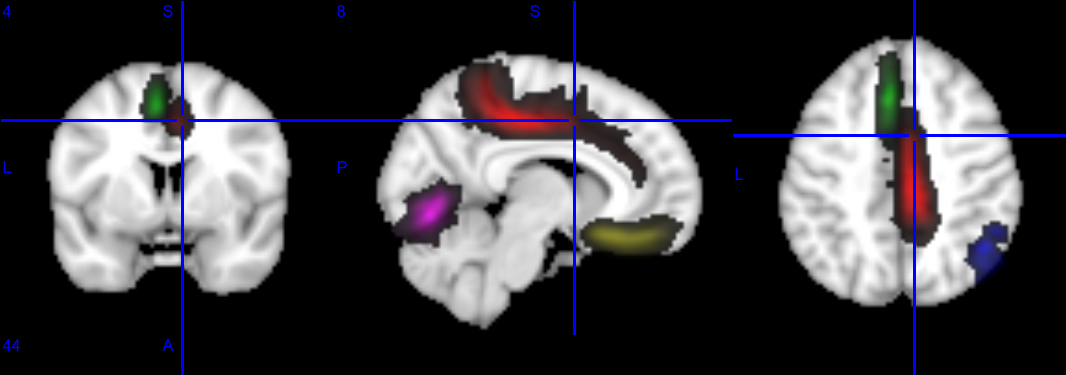
Probability maps
Probability maps corresponding to a few anatomical landmarks (cortical folds) overlaid on a T1 template
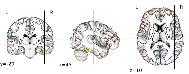
Outlines
The outline at 50% of all the structures
Labels location
Location of sulcal labels mapped on a 3D representation of a refined version of the anatomical atlas (after a sulcuswise non-linear registration scheme).
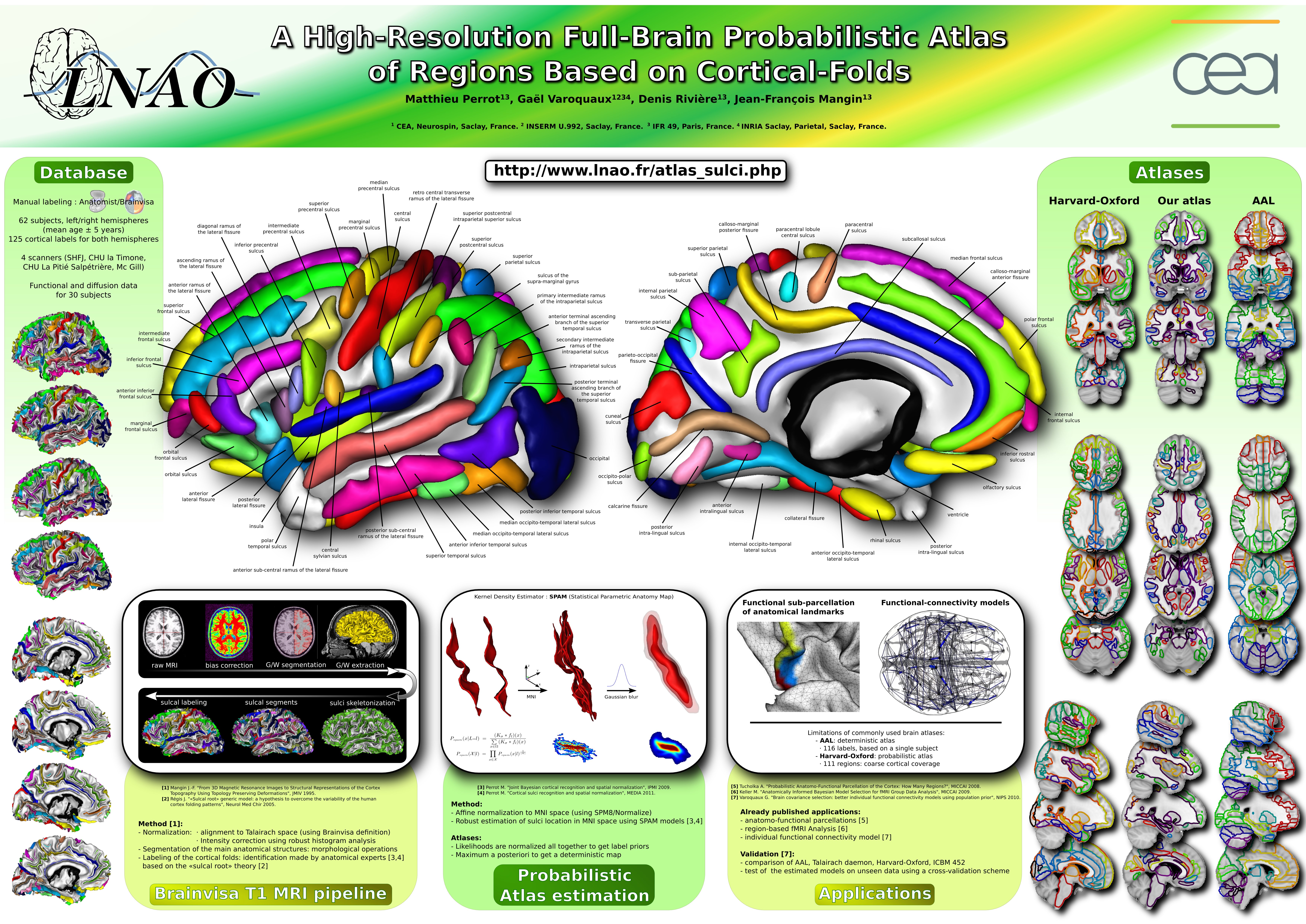
HBM 2011 poster
A high-resolution full-brain probabilistic atlas of regions based on cortical folds
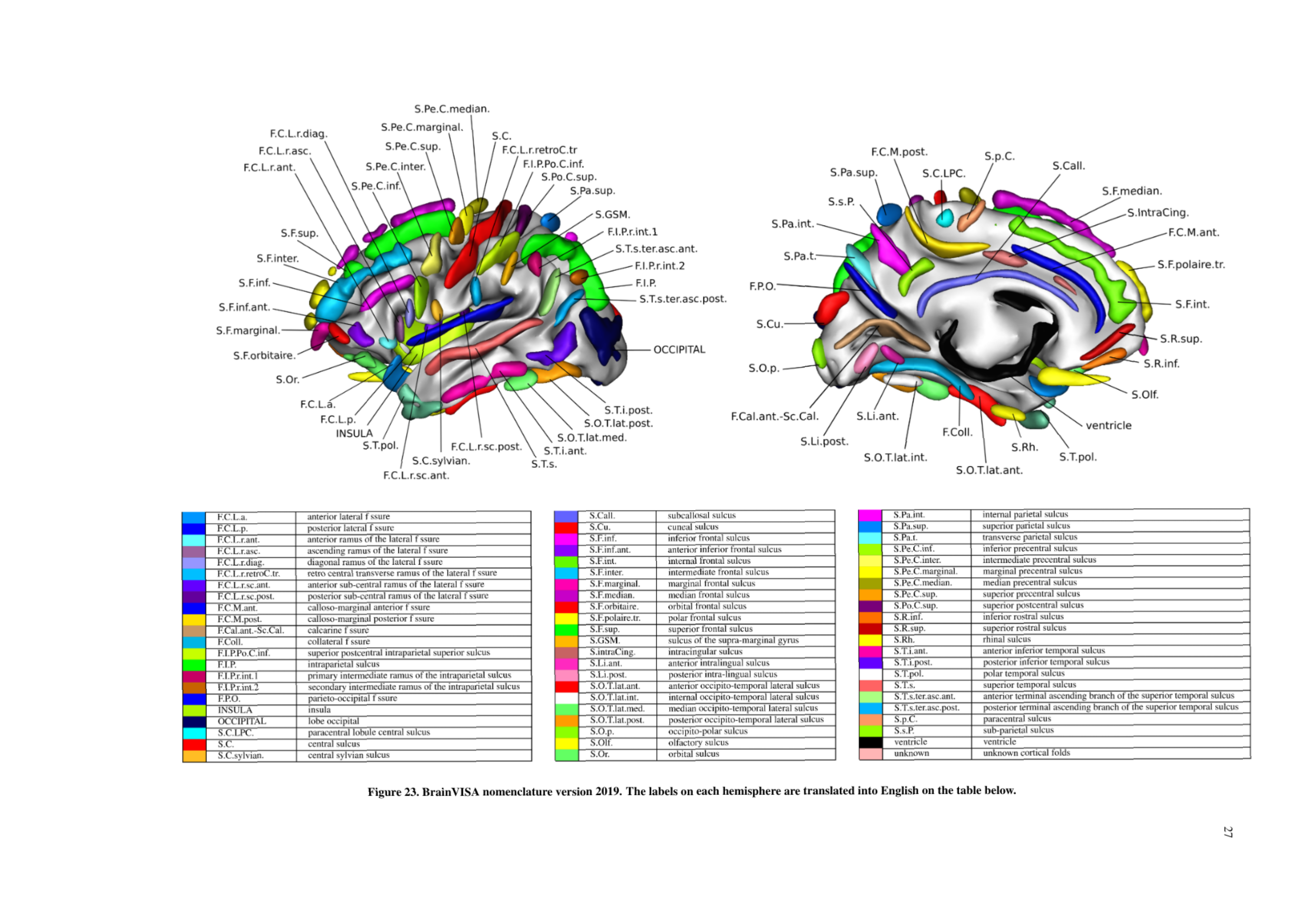
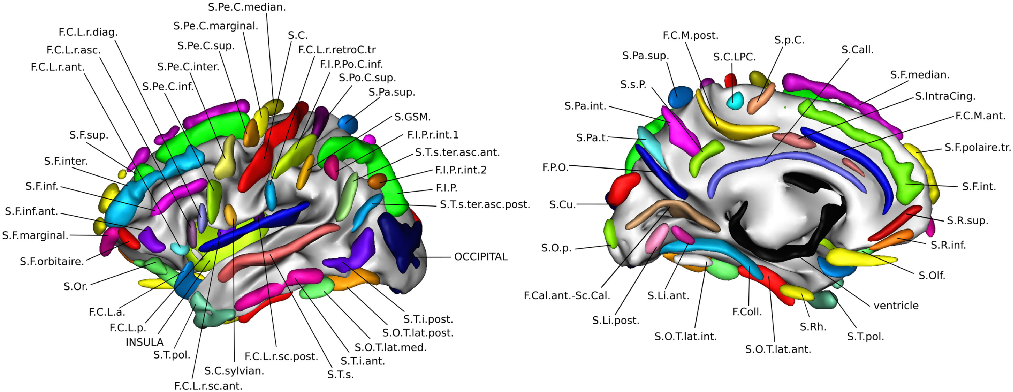
Atlas of Sulci with nomenclature table
Location of sulci named by their Brainvisa acronyms or their full anatomical names
Downloads
Download the Atlas (nii.gz file, 71Mo, in MNI space, 2mm isotropic voxels)
Table giving the names of the anatomical structures (csv file)
CSV of anatomical structures - fields : index, location (x,y,z), color(r,g,b), brainvisa acronym and anatomical label.
Table of sulcal labels - Meanings of Brainvisa acronyms used for sulcal structures
The older sulci labels used before BrainVisa/Morphologist 3.2, and handled by the older ANN models 2001. There are slight changes, but there are changes.
References
Atlas estimation
Perrot, D. Riviere, A. Tucholka, J.-F. Mangin : Joint Bayesian Cortical Sulci Recognition and Spatial Normalization, IPMI 2009 link bibtex-entry paper
Perrot, D. Rivière, and J.-F. Mangin. Cortical sulci recognition and spatial normalization. Medical Image Analysis 2011 link bibtex-entry paper
Applications
Defining anatomo-functional parcels
Alan Tucholka, Bertrand Thirion, Matthieu Perrot, Philippe Pinel, Jean-François Mangin and Jean-Baptiste Poline : Probabilistic Anatomo-Functional Parcellation of the Cortex : How Many Regions ?, MICCAI 2008 link bibtex-entry paper
Refining functional MRI analysis
Merlin Keller, Marc Lavielle, Matthieu Perrot and Alexis Roche. Anatomically Informed Bayesian Model Selection for fMRI Group Data Analysis. MICCAI 2009 link bibtex-entry paper
Functional connectivity
Gaël Varoquaux, Alexandre Gramfort, Jean-Baptiste Poline, Bertrand Thirion. Brain covariance selection : better individual functional connectivity models using population prior. NIPS 2010 paper
