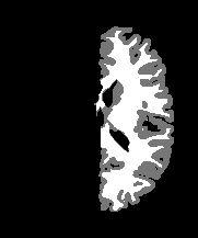


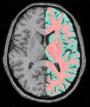
Grey/white classification for "Voxel Based Morphetry"
This procedure yields a grey/white classification of each hemisphere for people wishing to test the influence of classification algorithms (SPM, MNI's tools, SHFJ's tools...) on the final VBM result.
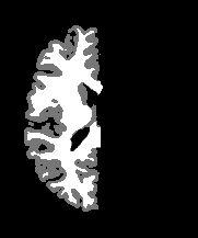
The proposed method deals with each hemisphere individually, in order to prevent mixing between the two internal faces or between low cortex faces and cerebellum, during the spatial smoothing performed before statistics.
Another feature of the proposed approach: we have decided to discard cerebellum because partial volume effects are in our opinion too important to reach a correct binary classification (a fuzzy classification might overcome this problem). Nevertheless, a classification of cerebellum or any other masked area can be obtained from the command line VipGreyWhiteClassification.
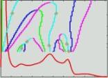
The classification algorithm relies mainly on the result of the histogram analysis yielded by Vip Histogram Analysis. The result does not stem from a threshold but from a "Markovian regularisation": a voxel with intermediate grey level is attributed to the majority class in the surrounding.
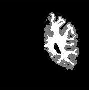
A result example:
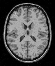

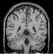
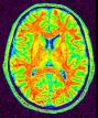
mri_corrected: T1 MRI Bias Corrected ( input )
histo_analysis: Histo Analysis ( input )
split_mask: Split Brain Mask ( input )
Side: Choice ( input )
left_grey_white: Left Grey White Mask ( output )
right_grey_white: Right Grey White Mask ( output )classification (grey=100,white=200)
Toolbox : Morphologist
User level : 2
Identifier :
AnaComputeHemiGreyWhiteClassifSupported file formats :
mri_corrected :gz compressed NIFTI-1 image, Aperio svs, BMP image, DICOM image, Directory, ECAT i image, ECAT v image, FDF image, FreesurferMGH, FreesurferMGZ, GIF image, GIS image, Hamamatsu ndpi, Hamamatsu vms, Hamamatsu vmu, JPEG image, Leica scn, MINC image, NIFTI-1 image, PBM image, PGM image, PNG image, PPM image, SPM image, Sakura svslide, TIFF image, TIFF image, TIFF(.tif) image, TIFF(.tif) image, VIDA image, Ventana bif, XBM image, XPM image, Zeiss czi, gz compressed MINC image, gz compressed NIFTI-1 imagehisto_analysis :Histo Analysis, Histo Analysissplit_mask :gz compressed NIFTI-1 image, Aperio svs, BMP image, DICOM image, Directory, ECAT i image, ECAT v image, FDF image, FreesurferMGH, FreesurferMGZ, GIF image, GIS image, Hamamatsu ndpi, Hamamatsu vms, Hamamatsu vmu, JPEG image, Leica scn, MINC image, NIFTI-1 image, PBM image, PGM image, PNG image, PPM image, SPM image, Sakura svslide, TIFF image, TIFF image, TIFF(.tif) image, TIFF(.tif) image, VIDA image, Ventana bif, XBM image, XPM image, Zeiss czi, gz compressed MINC image, gz compressed NIFTI-1 imageleft_grey_white :gz compressed NIFTI-1 image, BMP image, DICOM image, Directory, ECAT i image, ECAT v image, FDF image, GIF image, GIS image, JPEG image, MINC image, NIFTI-1 image, PBM image, PGM image, PNG image, PPM image, SPM image, TIFF image, TIFF(.tif) image, VIDA image, XBM image, XPM image, gz compressed MINC image, gz compressed NIFTI-1 imageright_grey_white :gz compressed NIFTI-1 image, BMP image, DICOM image, Directory, ECAT i image, ECAT v image, FDF image, GIF image, GIS image, JPEG image, MINC image, NIFTI-1 image, PBM image, PGM image, PNG image, PPM image, SPM image, TIFF image, TIFF(.tif) image, VIDA image, XBM image, XPM image, gz compressed MINC image, gz compressed NIFTI-1 image